La rate humaine. Tuberculose — Photo

La rate humaine. Tuberculose — Image libre de droits

Micrographie photonique d'une rate d'une personne atteinte de tuberculose avancée. Plusieurs granulomes typiques avec une nécrose centrale (nécrose caséeuse) peuvent être observés .

 — Image de jlcalvo@ucm.es

Même série:

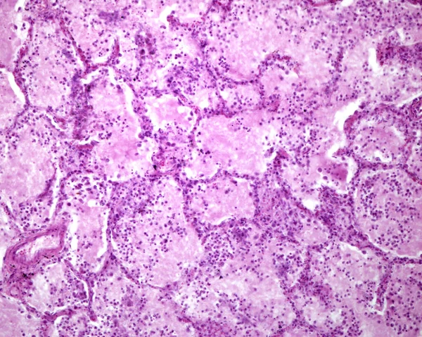
Histological section of a human lung affected by an acute pneumonia. The lumen of alveoli is occupied by a liquid of oedema which contains acute inflammatory infiltrates (with predominance of neutrophil granulocytes).
Histological Section Human Lung Affected Acute Pneumonia Lumen Alveoli Occupied — Photo
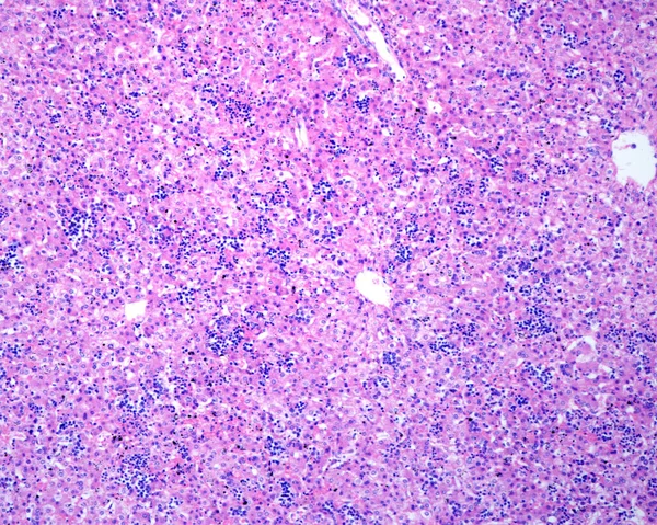
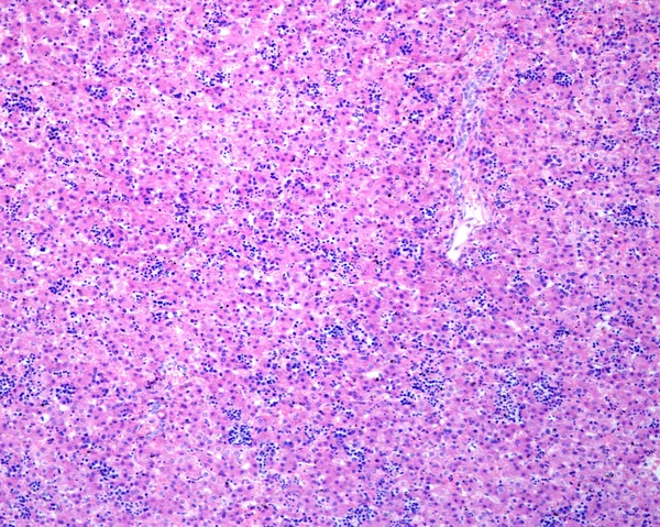
Liver. Erythroblastosis fetalis is characterized by intravascular proliferation of immature precursors of erythrocytes. Histologically, accumulation of erythroblasts in blood sinusoids is observed
Liver Erythroblastosis Fetalis Characterized Intravascular Proliferation Immature Precursors Erythrocytes Histologically — Photo
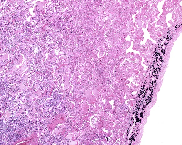
Periphery of a human lung affected by an acute pneumonia. The lumen of alveoli is occupied by liquid of oedema which contains acute inflammatory infiltrates (with predominance of neutrophil granulocytes). On the right, the pleura shows many carbon-lo
Periphery Human Lung Affected Acute Pneumonia Lumen Alveoli Occupied Liquid — Photo
Human spleen amyloidosis. Amyloidosis is caused by abnormal folding of normal soluble proteins leading to fibril formation. These clumps of protein are called amyloid deposits. The spleen is involved in most cases of systemic amyloidosis. The microgr
Human Spleen Amyloidosis Amyloidosis Caused Abnormal Folding Normal Soluble Proteins — Photo
Neurone coloré avec du violet crésyle montrant un corps de Barr (chromatine sexuelle) situé près d'un gros nucléole. Microscope optique .
Corps de Barr. Neurone — Photo
Human liver. Drug-induced liver injury by carbon tetrachloride. Low magnification micrograph showing several clear areas of degeneration and necrosis of hepatocytes with little or no inflammation.
Human Liver Drug Induced Liver Injury Carbon Tetrachloride Low Magnification — Photo
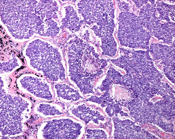
Human lung. Small cell carcinoma. Clusters and sheets of small cells, round to oval to spindle-shaped, finely granular chromatin, and very scant cytoplasm, numerous mitotic figures, and central areas of necrosis.
Human Lung Small Cell Carcinoma Clusters Sheets Small Cells Oval — Photo
Faible grossissement d'une prostate humaine chez un homme de 70 ans. La prostate apparaît avec des alvéoles dilatées, qui contiennent de nombreux corps amylacés (concrétions prostatiques) dans leur lumière. Microscope photonique. Tache d'hématoxyline et d'éosine .
La prostate humaine. Corpus amylacées — Photo
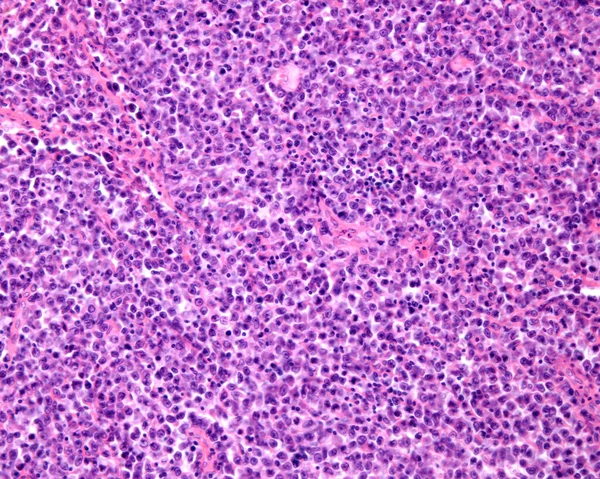
Reticulosarcoma. Light micrograph from a lymph node in a case of reticulosarcoma, a malignant lymphoma type. The normal structure of the lymph node is totally occupied by sarcoma malignant cells.
Reticulosarcoma Light Micrograph Lymph Node Case Reticulosarcoma Malignant Lymphoma Type — Photo
Human spleen amyloidosis. Amyloidosis is caused by abnormal folding of normal soluble proteins leading to fibril formation. These clumps of protein are called amyloid deposits. The spleen is involved in most cases of systemic amyloidosis. The microgr
Human Spleen Amyloidosis Amyloidosis Caused Abnormal Folding Normal Soluble Proteins — Photo
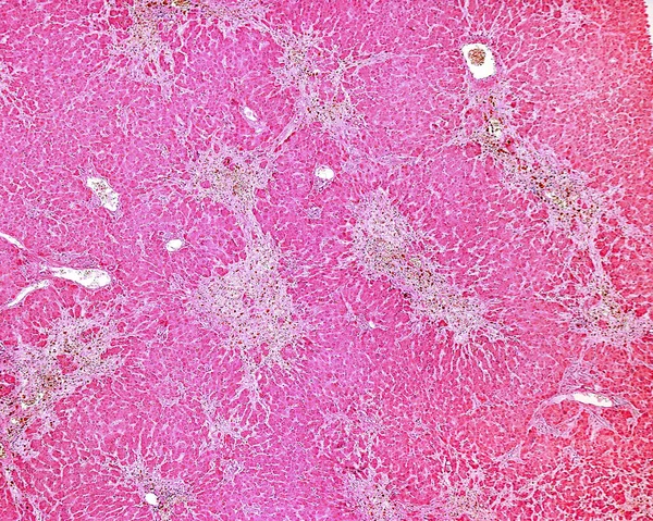
Human liver. Cirrhosis. Low magnification micrograph showing regenerating nodules of hepatocytes (with an extensive fatty change), separated by fibrous septa with chronic inflammatory infiltrates. Cirrhosis of the liver is a result of severe damage t
Human Liver Cirrhosis Low Magnification Micrograph Showing Regenerating Nodules Hepatocytes — Photo
Human lymph node. Metastasis of a malignant melanoma. The normal lymphoid tissue of lymph node has completely disappeared and has been replaced by malignant melanoma pigmented and non-pigmented cells.
Human Lymph Node Metastasis Malignant Melanoma Normal Lymphoid Tissue Lymph — Photo
Human spleen amyloidosis. Amyloidosis is caused by abnormal folding of normal soluble proteins leading to fibril formation. These clumps of protein are called amyloid deposits. The spleen is involved in most cases of systemic amyloidosis. The microgr
Human Spleen Amyloidosis Amyloidosis Caused Abnormal Folding Normal Soluble Proteins — Photo
Liver. Erythroblastosis fetalis is characterized by intravascular proliferation of immature precursors of erythrocytes. Histologically, accumulation of erythroblasts in blood sinusoids is observed
Liver Erythroblastosis Fetalis Characterized Intravascular Proliferation Immature Precursors Erythrocytes Histologically — Photo
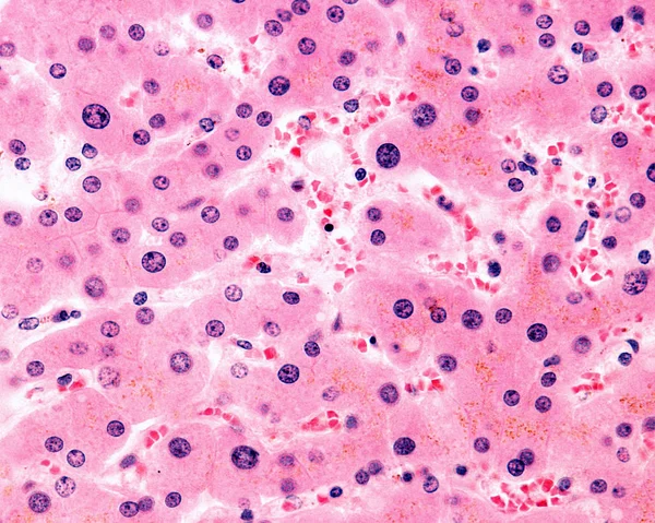
Hépatocytes polyploïdes. Plusieurs hépatocytes montrent un noyau très grand. Ces cellules sont polyploïdes, avec 4, 8, 16 fois ou plus le complément chromosomique haploïde. Les hépatocytes contiennent d'abondantes granules de pigment biliaire .
Noyaux polyploïdes — Photo

Informations d'utilisation

Vous pouvez utiliser cette photo libre de droits "La rate humaine. Tuberculose" à des fins personnelles et commerciales conformément à la licence Standard ou Étendue. La licence Standard couvre la plupart des cas d’utilisation, comprenant la publicité, les conceptions d’interface utilisateur et l’emballage de produits, et permet jusqu’à 500 000 copies imprimées. La licence Étendue autorise tous les cas d’utilisation sous la licence Standard avec des droits d’impression illimités et vous permet d’utiliser les images téléchargées pour la vente de marchandise, la revente de produits ou la distribution gratuite.

Vous pouvez acheter cette photo et la télécharger en haute définition jusqu’à 3840x3072. Date de l’upload: 14 janv. 2018